The advantage of digital photography over molding in dental implant placement

The advantage of digital photography over molding in dental implant placement

History of traditional molding and digital photography
Digital imaging or imaging systems have been available for over 25 years but have recently become popular. The first scanable implant was used in 2004, which used coded heel abutment. This abutment provided the dentist with all the three-dimensional information about the position of the dental implant relative to the adjacent and opposite teeth.
In this digital method, as in normal molding, the exact position of the dental implant, including the internal position of the anti-rotational properties such as anti-rotational grooves and internal hexagon, was recorded. Most of these scans were performed after normal tooth molding, and the model obtained by the laboratory was scanned. This technique is still used, although it is not justified in terms of time and cost.
In 2010, a scanable molding coupling was introduced to the market, which was placed on the implant and scanned inside the mouth using a parallel focus imaging system. Although this eliminated the need for conventional paste molding, this approach was limited to an implant and an intraoral scan.
In the last few years, a number of scanners have entered the market that can scan multiple implants simultaneously.
The two main types of scanners used today are: 1) the type of blue LED (light emitting diode) which is an optical scanner that usually requires powder to work with and 2) scanners that use lasers to scan They record the distance from the tooth surface and build a digital model based on it.
Recently, most scanners use laser technology to eliminate the need to use powder. The need to use powder in type I scanners is one of the first disadvantages of this method for dentists.
Dental implant imaging method
In the conventional method, implants, bridges, and veneers must be molded and modeled. In ordinary repairs, the molding is taken with polyester or polyvinyl siloxane and then the original model is made with special gypsum materials.
After making the model, the template is indexed accordingly. This process is time consuming. But digital intraoral imaging takes about three minutes less than conventional molding from the time the cap is removed to the time it is replaced.
After the molding is done with the usual method, the model must be made. In conventional molding, the mold must be cleaned and sent to a dental laboratory to make a model. In most cases, the technician will not even check the submitted physical model within 24 to 72 hours.
In the digital photography method, which is done using an oral camera or scanner, all important data is sent over the Internet in the form of digital files. In this way, you can start designing the abutment and implant cover on the same day of digital molding without having to make their model.
Although there are many types of digital intraoral molding machines on the market, they all have the same function. The first step immediately after the implant is fused to the bone (the process of bone integration) and the tissues heal, is to ask the patient to go to the office for a digital scan of the implant.
This scan is similar in performance to conventional molding, but instead of the usual implant coupling, a scanable coupling is used.
After the scanable coupling is placed on the implant, the radiographic image confirms its correct position. This step is sensitive because the final abutment and veneer are to be made and used based on it.
No dentist wants to impose the cost of rebuilding the restoration on himself or the patient, so accurate abutments must be scanned and made.
After that, the scan is performed and a three-dimensional model of this part of the mouth is displayed on the screen, which includes all the necessary surfaces of the scanable molding coupling to make the abutment and veneer.
These surfaces include molding cups, the contact areas of all adjacent teeth, gingival tissue, anterior dental arch, and bite.
After the scan, the laboratory instructions, including the color, the choice of abutment material (zirconia or titanium) and the material of the veneer, are determined and sent to the repair center. Sending is done via the Internet.
At this stage the model, abutment and veneer may be made by a laboratory center or performed in separate centers. It depends on the scanning system used.
After making the abutment and veneer, the patient comes to install the restoration on the implant. This step and the steps after it are no different from the usual method.
The benefits of digital photography for implant placement
One of the most important advantages of this method is that it does not require molding materials, so problems such as unpleasant taste of molding materials and the feeling of gurgling are not experienced in the patient.
Experts are well aware that patients hate the molding process and may even be so afraid of it that they avoid going to the dentist.
In a recent study, patients with no previous history of conventional and digital casting all preferred digital casting.
The findings of this study confirm the need to develop digital solutions in the formulation and fabrication of restorations.
Patients' personal preferences should not determine the treatment method, but all aspects of work should be considered. The main concern of dentists for choosing new technology or techniques is whether the new method has the accuracy, speed, convenience, and predictability of the previous methods, and whether they are as cost-effective as they are.
It is difficult to achieve technology that provides all of these features at a higher level, and usually some less important features fall victim to the more important features.
Accuracy in dental implant restoration is a key factor in the beauty and longevity of treatment results. Restorations that do not work with high accuracy will ultimately fail.
In the conventional molding method, there are many steps and in each step, a percentage of error must be considered, so in general, the probability of error in this method is higher.
As mentioned in the conventional molding method, the implant is scanned based on the gypsum mold sent to the laboratory, so any errors that occur in the initial molding are added to the final repair and the errors associated with the subsequent steps.
The length of time a patient has to sit in a dental chair is a determining factor in the cost of treatment. Numerous studies have shown that in a digital molding system, the length of time a patient sits in a dental chair for molding is shorter.
In addition, the number of times a patient has to see a dentist or dentist is almost zero because everything is done digitally and requires an individual presence.
After a three-dimensional model is made in the laboratory, it is evaluated for tooth contact and implant position before making an abutment or veneer. If there is a problem in the model, it is identified before making the restoration, thus saving time, cost and energy of the patient and the dentist.
In the digital method, work can be done without a model, so the need to build a model and a tray and the associated costs are avoided.
The only cost that is higher in the digital method than the usual method is the cost of buying a scanner, but this cost will ultimately be economically justified given the above.
The Advantage of Digital Imaging Instead of Molding in Dental Implant Implantation
Computer-assisted implant placement
Nowadays, digital implant implants (code / low) have found their place in the dental world. The 3D scanner (camera) inside the mouth has revolutionized the process of imaging and molding the mouth. However, many specialists have focused only on the restorative aspects of this new technology, while this advanced technology can be used for dental implants.
Ordinary tooth molding
In the typical molding process taught at the School of Dentistry, molding paste is used. The first step is to choose the right molding material (polyvinyl or alginate) and mixing pad, spatula, glue, lubricant, mixer and trays in different sizes.
Then the most suitable in the maxilla (maxillary) and mandible (mandibular) should be selected and the materials should be combined. Imagine doing this in front of a patient and not at all interesting for many patients who are afraid of dentistry.
Some people sneeze when they get mold in their mouth and can't stand it. Molding is a stressful task for both the patient and the technician.
If all goes well, only one molding is needed, but if the slightest mistake occurs, whether it is related to laboratory work or anything else, this undesirable process must be repeated.
Fortunately, digital molding has greatly facilitated this process. The work is simply done with a special scanner or camera and the image is displayed on the screen. All you have to do is disconnect the scanner from the laptop and connect it to the USB port of the computer you are using.
A collection of digital images inside the mouth is taken very quickly and processed by a computer. The whole process of digital molding is done in a few minutes. The speed, accuracy and efficiency of digital scanners have made many patients and dentists prefer this method.
The manual part of this device is light and can be moved easily. Once the digital file is ready, it is sent to a laboratory or dentist or opened in the implant surgery surgery planning software.
Implant implant surgery guide
When placing a dental implant, it is always advisable to use a surgical guide. In addition to helping to place the implant correctly, this approach ensures that the correct position is chosen and the distance between the teeth is observed.
In this way, the need for a custom abutment during the implant installation repair phase may be eliminated and treatment costs may be reduced.
Making a surgical guide with conventional molding requires the use of polyvinyl or alginate and pouring the material in two steps. After the paste has hardened, an artificial tooth is placed in the toothless area of the model and made by heating the thermoplastic thermocouple.
Fortunately, with a digital scanner, as soon as the oral scan is done, the digital file is sent to the lab to have a virtual tooth added, and a guide is made based on it and sent to the office. So in this method, no model is made, so additional costs are avoided.
Dental implant placement
After placing the implant and welding it to the jawbone (bone integrity), it must be determined what type of abutment is needed. You can choose standard abutment or stock (prefabricated) or custom abutment (which is made especially for you).
The abutment is the interface between the base of the implant and the veneer. The standard abutment is made by the implant manufacturer and is usually used when there is no concern about the angle of the implant. A custom abutment is selected when its direction, position, or relationship to other teeth are specific and different.
If the molding is done with paste and it is used for standard abutment, the molding coupling is placed on the abutment. Viscous material is injected into the patient's mouth and mouth. Once it has taken hold and hardened, it comes out of the mouth with the coupling. The analog of the implant is placed in the coping and the laboratory receives it and makes a stone model of it.
If a custom abutment is to be made in the same way as paste molding, the implant will be molded from the top of the implant fixture and an abutment will be made based on it for the patient. In coupling molding level with the top of the implant fixture, the same molding technique is performed.
Due to the many steps in this molding method, the probability of error increases. In the digital method, whether you want to create a standard abutment or a custom one, a criterion can be used. Digital molding is done with higher accuracy and is instant.
In the picture below, the patient has three tooth gaps and several implants are to be placed for him. The standard abutment was placed in the jaw and CS 3500 was used for molding.
There is no need to spend time to make more and the paste is not placed in the patient's mouth so it saves time and the patient is more comfortable.
Immediate dental implants
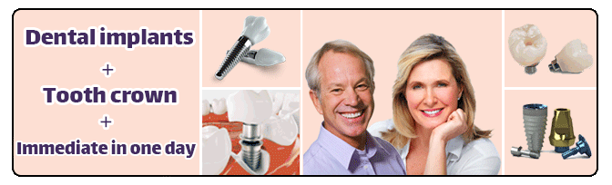
Dental implants + Tooth crown + immediate in one day
A Dental Implant or fixture replaces a lost tooth the size of a tooth root.
After the implant is welded, the casting is done and after the prosthesis is made in the laboratory, the crown is delivered to the patient; Sometimes this process can be done even in one day.
If you have lost one or more of your teeth and need more information,
So contact us as soon as possible to schedule a check-up and consultation.